
When diagnosing and treating patients, it is important to use the correct technology. MRI and MRA scans are very similar, but they differ in what they are used for. In both cases powerful magnetic field, radio waves and a computer to produce detailed pictures of the body. However, what they examine is different for each scan.
MRI
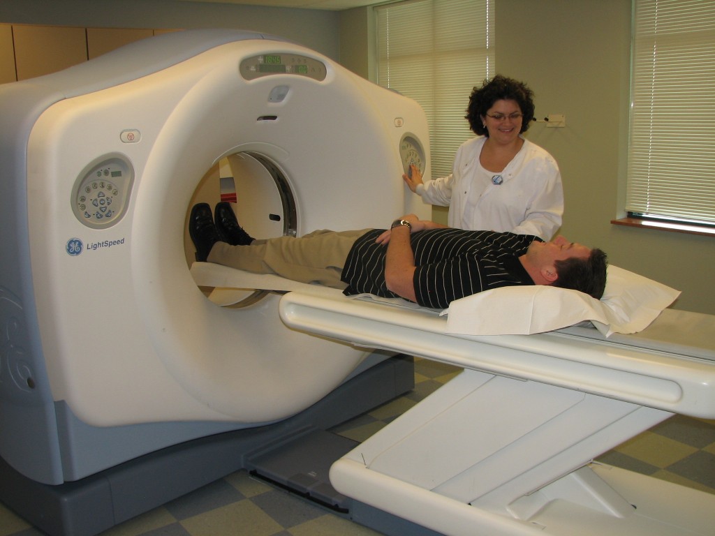
An MRI, or magnetic resonance imaging, is the technology behind an MRA, and it is used to examine soft ligament tissues and tendon injuries. MRI scans can be used to discover, treat, and monitor disease. It is a non-invasive, painless medical test that is very good at looking at the anatomy of muscles, tendons, ligaments, and internal organs, such as the liver, kidneys, heart, brain, and spine. It can also assist with surgical planning. When using the MRI scanner, the radiologist or doctor can see very detailed images of almost all the tissue in the body. It is even possible to gather data about the different types of tissues by changing the timing of the radio wave pulses.
MRI Scans can be used for:
- To find aneurysms.
- Evaluate bone and joint disorders and locate bone infections or tumors.
- Understand disorders of the eye and inner ear.
- To monitor multiple sclerosis.
- To discover or help treat spinal cord injuries.
- To see if a patient has had a stroke.
- To locate tumors or cancers inside many of the body’s organs.
- To help detect breast cancer in women who have dense breast tissue or who are at high risk of the disease.
- To assess heart function, such as the size and health of heart chambers, thickness, and movement of heart walls, the extent of damage caused by a heart attack or heart disease, structural problems in the aorta, and/or inflammation or blockages in heart blood vessels.
MRA
A Magnetic Resonance Anigram (or an MRA) is a type of MRI scan that uses MRI’s magnetic fields and radio waves to produce pictures of blood vessels inside the body. This non-invasive, painless medical test helps physicians locate problems that may cause reduced blood flow. For MRA’s, both the blood flow and the condition of the blood vessel walls can be seen. MRA scans are well suited to examine arteries in the brain, neck, chest, and abdomen. During MRA scans, the area of the body being studied is put inside an MRI machine. Contrast material (a dye) is used to make the blood vessels show up more clearly.
MRI Scans can be used for:
- Finding bulges (aneurysms), clots, or build-ups of fat and calcium deposits in the blood vessels leading to the brain.
- To find aneurysms or tears in the aorta, which carries blood from the heart to the rest of the body.
- To find blood clots in arteries and veins.
- To see if any blood vessels leading to the heart, lungs, kidneys, or legs are narrowing. Narrowing blood vessels can lead to hypertension, painful walking, and non-healing ulcers.
Seton Imaging is one of the areas longest-established Radiology practices. We are proud to offer our patients a complete range of diagnostic imaging services and procedures. Our experienced and skilled team of radiologists work closely with your referring physician to bring you the best in diagnostic imaging services and the absolute highest quality of care.